1. Ankle joint AP(AnteroPosterior) Stress View
촬영 목적 Varus(or inversion: 안쪽 번짐) stress view= tear of lateral collateral ligament(가쪽 곁 인대의 찢어짐)와 ankle joint instability(발목관절의 불안정)시 시행 / Valgus(or eversion: 가쪽 번짐) stress view= tear of anterior talofibular ligament(앞 목말종아리 인대의 찢어짐)와 ankle joint instability시 시행, deep injury of deltoid ligament(세모인대의 깊은 상처)일 때 시행 한다. Film size 는 8x10 half crosswise를 사용하고 촬영 table에 설치되어 있는 toles device위에 knee joint를 extension 검사측 ankle joint를 counter support를 위치시켜 발이 움직이 않게 한다. pressing 위치는 medial or lateral malleolus 위 5cm에 15daN의 압력으로 pressing 한다. 평가 기준은 ligament injury시 검사하는 varus stress는 ankle joint의 경사가 25도~30도 이거나 정상 ankle joint의 8도~10도 이상이 되는지에 따라 joint space가 차이가 난다. 또 valgus stress 시에는 medial articular space(안쪽 관절의 공간)이 3cm 이상이 되는지 유무를 관찰해야 한다.

2. Knee joint AP(AnteroPosterior) Projection=무릎관절(슬관절) 앞뒤방향 촬영
촬영목적은 medial and lateral condyle fracture of femur(넙다리뼈의 안쪽과 가쪽 관절융기 골절), medial and lateral intercondyloid eminence fracture of tibia(정강뼈의 안쪽과 가쪽 관절융기사이 융기골절), fracture of tibial spine and proximal fibula(정강뼈 가시의 골절 및 몸쪽 종아리뼈의 골절), compartment of medial and lateral joint(안쪽과 가쪽 관절의 구획), osteochondral fracture(골 연골의 골절) 및 spontaneous osteonecrosis(자연발생적인 골 괴사)의 관찰 등이 있다. Film size는 10x12 half crosswise를 사용하고, 촬영 table 위에서 knee joint를 extenstion시켜서 앉거나 똑바로 눕게 한 후 머리에 베개를 받쳐주고, pelvis가 회전 되지 않도록 한다. object center는 patella apex(무릎뼈 끝) 아래 1cm 지점에 준다. 평가기준으로는 epicondyle of femur와 condyle of tibia의 joint space가 대칭적으로 나타나야 한다.

1.넙다리뼈(대퇴골,femur) 2.무릎뼈(슬개골,patella)
3.융기사이오목(과간와,intercondylar fossa) 4.안쪽위관절융기(내측상과,medial epicondyle)
5.안쪽관절융기(내측과,medial condyle) 6.가쪽위관절융기(외측상과,lateral epicondyle)
7.가쪽관절융기(외측과,lateral condyle) 8.관절 융기 사이 융기(inter-condyloid eminence or tibial plateau)
9.정강뼈(경골,tibia) 10.종아리뼈(비골,fibula)
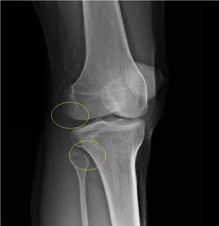
3. Knee joint Medial Oblique Projection=무릎관절(슬관절) 안쪽 사방향 촬영
촬영목적은 proximal tibiofibular joint의 관찰, lateral condyle fracture of femur and tibia 등을 볼 수 있다. Part position은 long axis of leg를 film mid line에 일치시키고 leg를 안쪽으로 45도 회전시킨다.(intercondylar line이 film면에 45도) 영상기준은 lateral condyle of femur and tibia는 반측면상으로 나타나야 한다.
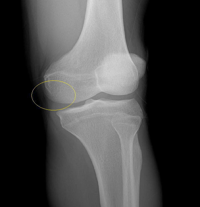
4. Knee joint Lateral Oblique Projection=무릎관절(슬관절) 가쪽 사방향 촬영
촬영목적은 medial condyle fracture of femur and tibia 이다. Part position은 long axis of leg를 film mid line에 일치시키고 leg를 가쪽으로 45도 회전시킨다.(intercondylar line이 film면에 45도) 영상기준은 medial condyle of femur and tibia는 반측면상으로 나타나야 한다.
'지식을 쌓아가는 일' 카테고리의 다른 글
| Knee PA Axial, Patella AP or PA Tangential (0) | 2021.12.06 |
|---|---|
| Knee LAT,Weight bearing AP, Stress view (0) | 2021.12.05 |
| Clavicle AP,Axial / AC joint (0) | 2021.12.03 |
| Foot AP,OBL,LAT (0) | 2021.12.02 |
| Shoulder Axial, Hill-Sachs , Stryker Notch View (0) | 2021.12.01 |


댓글