1. Elbow joint AP(AnteroPosterior) projection=팔꿉관절(주관절) 전후방향 촬영
촬영목적은 supracondyle, transcondyle, and intercondyle fracture of distal humerus(먼쪽 위팔뼈의 위 관절면, 수평관절면 및 내측 관절면 골절)medial and lateral epicondyle fracture of the humerus(위팔뼈의 안쪽과 가쪽 위 관절융기 골절)varus and valgus deformity, fracture of capitulum and trochlear of the humerus(위팔뼈의 작은 머리와 도르래 골절), lateral fracture of radial head이다. Film size는 8"x10" half crosswise를 사용하고 sitting이나 erect한 상태에서 long axis of forearm을 film midline에 일치시키고, both epicondyle of humerus가 film 면에 평행돠게 조정한다. object center는 mid-point of elbow joint에 준다. 촬영 point는 몸쪽노자관절(상요척관절)이 넓게 촬영되어야 한다. humerus 등쪽을 밀착시키기 위해 의자를 낮게 하는 등의 방법을 취하고, 관절간극을 선명하게 나타내기 위해 최대한 elbow를 extention 하고, humerus와 forearm이 cassette면에 같은 각도가 되도록 고정한다. 이 경우 forearm의 아래에 보조구를 놓아 고정하든지 검사반대측 팔도 검사측 손목을 고정하여 움직임을 방지하면 좋다. 환부가 명확한 경우는 그 부위를 중심으로 촬영한다. epicondyle 선의 각도의 계측에 의해 elbow의 굴곡각에 관계없이 끝 부분 골편의 경사 상태를 파악할 수 있다. humerus 축과 forearm 축이 이루는 각(Carring 각)은 팔꿈치를 펴고 supination을 했을 때에는 15도 정도의 외반이 정상이다.

1.위팔뼈(상완골,humerus) 2.팔꿈치돌기(주두돌기,olecranon process)
3.팔꿈치오목(주두와,olecranon fossa) 4.안쪽위관절융기(내측상과,medial epicondyle)
5.가쪽위관절융기(외측상과,lateal epicondyle) 6.갈고리돌기(구상돌기,coronoid process)
7.위팔뼈작은머리(상완골소두,capitulum of humerus) 8. 노뼈거친면(요골조면, radial tuberosity)
9.자뼈(척골,ulna) 10.노뼈(요골,radius)
2. Elbow joint Latreal projection=팔꿉관절(주관절) 측방향 촬영
촬영목적은 distal humerus와 proximal forearm의 측면 상이 나타난다. supracondyle and intercondyle fracture of distal humerus(먼쪽 위팔뼈의 위 관절면과 안쪽 관절면 내의 골절),anterior fracture and dislocation of radial head(노뼈 머리의 앞쪽 골절과 탈구),fracture of olecranon process and dislocation of elbow joint(팔꿈치 머리돌기의 골절과 팔꿉 관절의 탈구),fat pad sign 등이 있다. Film size는 8"x10" half crosswise를 사용하고 sitting이나 erect한 상태에서 Long axis of forearm을 film midline에 일치시킨다. elbow joint를 90도 flexion을 시키고, both epicondyle of distal humerus와 both styloid process of forearm에 준다. object center는 mid-shaft of forearm에 준다. 촬영point는 trochlear sulcus of humerus(위팔뼈의 도르래 고랑), ridge of capitulum and trochlear(작은 머리와 도르래 능선)과 trochlear notch of ulna의 형태가 동심원 형태로 나타나야 한다. 팔꿈치 90도 lateral flexion 영상에서는 coronoid process 상전방의 지방이 관절액의 증가와 함께 그 하연이 수평방향으로 촬영된다. 관절내골절의 진단(특히 소아*사춘기)에 유용한 후방의 지방(postrior fat pad sign)은 정상에서는 보이지 않지만 관절액의 증가 또는 출혈 등에 의해 후방으로 밀려 나면 촬영된다. elbow 주변 연부조직이 의심될 때에는 관절을 30~35도 정도 구부려야 한다.
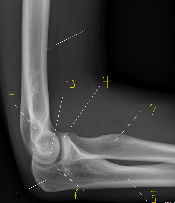
1.위팔뼈(상완골,humerus) 2.위관절융기(epicondyle)
3.도르래 고랑(trochlea sulcus) 4.갈고리돌기(구상돌기,coronoid process)
5.팔꿈치돌기(주두돌기,olecranon process) 6.도르래 패임(trochlea notch)
7.노뼈(요골,radius) 8.자뼈(척골,ulna)
'지식을 쌓아가는 일' 카테고리의 다른 글
| Shoulder Axial, Hill-Sachs , Stryker Notch View (0) | 2021.12.01 |
|---|---|
| Shoulder AP,OBL (0) | 2021.11.30 |
| Elbow OBL,Axial (0) | 2021.11.29 |
| Hand AP OBL, LAT (0) | 2021.11.26 |
| Hand PA ,OBL (0) | 2021.11.26 |


댓글